Ultrasound examination of females of the Black Sea bottlenose dolphin (Tursiops truncatus ponticus Barabash, 1940) during pregnancy
Semyonov V.A. 1, Danilova M.N. 1, Smyshnov A.V. 2, Osipova I.V.2
1 CJSC "Gelendzhik Dolphinarium", 130 Lunacharskogo St., Gelendzhik, 353460 Krasnodar Territory, Russian Federation
2 Municipal Health Institution "City Hospital", Gelendzhik, 353460 Krasnodar Territory, Russian Federation
Pregnancy is a critical period in the life of cetaceans, including those in captivity, related to the growth and development of the fetus.1 The use of ultrasound to monitor pregnancy in captive cetaceans provides valuable data on fetal morphology, development, and well-being, as well as on its measurements during pregnancy in female bottlenose dolphins, although these references need normative data.2,3,4 The purpose of these studies was to detect pregnancy in females of the Black Sea bottlenose dolphin (Tursiops truncatus ponticus Barabash, 1940) using ultrasound and to study the dynamics of the linear dimensions of the head and fetal chest, depending on its timing.
As a result of the research, we were able to determine pregnancy in females of the Black Sea bottlenose dolphin using ultrasound examination in the embryonic and fetal periods of its course, to identify dynamic differences in the size of the head and chest of the fetus in the period from the third to the twelfth months of pregnancy. If at the 2nd month of pregnancy (Fig. 1) we noted hyperechoic structures of the embryo in the chorion cavity, then at the 3rd month (Fig. 2) we already find the fetus and placenta. The embryo becomes a fetus with a baby-like configuration. The female is already in the fetal period of pregnancy, which is characterized by rapid growth of the fetus, differentiation of tissues, development of organs and systems from their rudiments, formation and formation of new functional systems that ensure the life of the fetus in the womb and the cub after birth. As can be seen from Table 1, the diameter of the fetal head at the 3rd and 4th months is still somewhat larger than the diameter of the developing chest, but at the 5th month the situation changes and until the 11th month the diameter of the chest is ahead of the diameter of the head. Then, at the 12th month, the measurements of these parts of the fetal body are compared again and reach almost 15 cm. Thus, it becomes clear that the use of the ultrasound diagnostic method to determine pregnancy in the Black Sea bottlenose dolphins is relevant and is the most reliable from the first months of its occurrence. Although the presented data were obtained by examining a relatively small number of pregnant individuals, at this stage of knowledge, they can probably be used as guidelines for determining the timing of pregnancy using ultrasound examination of Black Sea bottlenose dolphins.
Table 1. Dynamics of changes in the dorso-ventral dimensions of the head and chest (X ± m, cm) of the fetus during pregnancy in females of the Black Sea bottlenose dolphin
Month of pregnancy
Dorso-ventral dimensions of the head N=14, n=21
Dorso-ventral dimensions of the chest N=14, n=21
Number of studies X ± m, cm Number of studies X ± m, sm
3rd 1 3.04 1 2.68
4th 2 3.9±0.12 2 3.7±0.11
5th 2 4.3±0.11 2 5.3±0.01**
6th 4 5.4±0.25* 4 6.7±0.22**
7th 7 6.5±0.26* 7 7.7±0.20*
8th 8 8.1±0.42** 9 9.5±0.45**
9th 5 10.8±0.08 *** 5 11.4±0.18**
10th 6 10.9±0.29 6 12.7±0.11***
11th 7 12.0±0.20* 7 14.5±0.18
12th 4 14.9±0.54 ** 3 14.8±0.42
Total 45 - 45 -
Shortcuts:
X - arithmetic mean; m - standard error for the sample fraction;
N– the number of examined individuals; n– number of pregnancies studied
Significance of differences between this gestational age and the previous one: *- P < 0.05; ** – P< 0.01; *** – P< 0.001
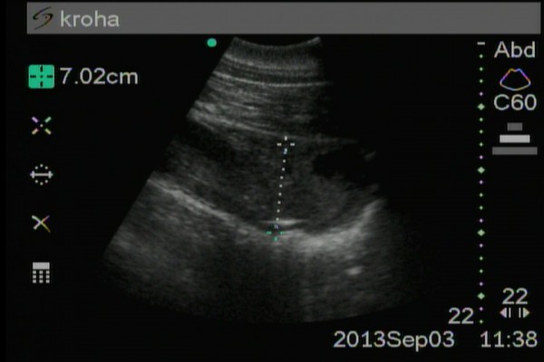
Fig.1 Second month of pregnancy
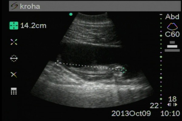
Fig.2 Third month of pregnancy
Thanks
The authors would like to express their gratitude for professional advice and practical assistance to the State Veterinary Administration of the Krasnodar Territory, State Budgetary Institution "Department of Veterinary Medicine of the City of Gelendzhik", Municipal Health Institution "City Hospital", Municipal Health Institution "Maternity Hospital".
Literature cited
1Robeck TR, Atkinson SKC, and Brook F. 2001. Reproduction. In: Dierauf LA, Gulland FMD, editors. CRC Handbook of Marine Mammal Medicine. second edition. Boca Raton. p 193-226.
2Brook F, Bonn WV, and Jensen ED. 2001. Ultrasonography. In: Dierauf
